Research

Dr. Federico has an active research program employing both functional and structural brain imaging of patients with epilepsy to better understand how focal seizures are generated and how they affect the brain. His lab pioneered intracranial EEG-fMRI and is the only Canadian centre performing these studies. His lab also pioneered the use of MRI and CT perfusion techniques to identify the seizure onset zone using post-ictal hypoperfusion.

His studies are primarily focused on the following three topic areas:

Intracranial EEG-fMRI

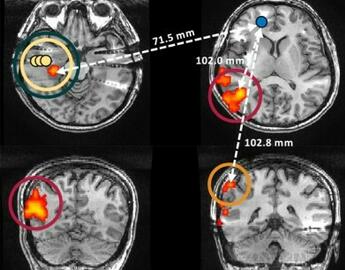
It is increasingly common for patients with drug-resistant epilepsy undergoing pre-surgical work-up to have electrodes implanted into their brain to more precisely identify the seizure-generating brain tissue (termed the seizure onset zone) for resection. The use of these intracranial electrodes over scalp electrodes greatly increases our ability to determine the location and extent of the seizure onset zone when the electrodes are placed nearby. However, if tissue distant to the electrodes is also involved in generating seizures, this tissue may be missed. The use of functional MRI (fMRI) concurrent with intracranial EEG overcomes this potential problem, allowing us to record from all locations within the brain, not just tissue immediately surrounding the electrodes, while maintaining the information from very fine timescales that intracranial EEG detects. Our lab is interested in using fMRI to record brain activation changes (BOLD response) throughout the brain at the time of epileptic discharges captured with the fine temporal resolution of EEG. We believe changes as the result of epileptic discharges may provide insight into regions of the brain responsible for creating seizures.

To date, we have studied the BOLD response to interictal discharges, and found concordance between areas identified using intracranial EEG-fMRI and those involved in generating spikes as well as the presumed seizure onset zone. Consequently, intracranial EEG-fMRI may be useful in defining the surgical target in epilepsy surgery.

Additionally, we are exploring the dynamic and static functional connectivity of regions associated with interictal spikes, in hopes that network dynamics may shed light on how epileptic activity alters the brain.

Click here to see our most recent paper on intracranial EEG-fMRI.

High Frequency Oscillations

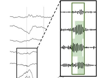
High frequency oscillations (HFOs), as the name suggests, are periods in time where the EEG signal becomes transiently high frequency (80-250Hz), or very high frequency (> 500Hz), resulting in an event resembling a ‘ripple’ in the recording. Although HFOs are produced physiologically in healthy individuals, they may also be produced under pathological conditions such as epilepsy. It is expected that physiological or healthy HFOs would be produced in regions throughout the brain, however pathological HFOs would be produced only in the regions of the brain that are important for seizure generation. Therefore, if pathological HFOs could be isolated, the regions that generated them would be of clinical importance.

High frequency oscillations are most easily detected using intracranial EEG, and we have begun investigating HFOs captured during our simultaneous intracranial EEG-fMRI studies. This will allow us to explore the hemodynamic response to HFOs, and further explore the relationship between HFOs and epilepsy, especially as it relates to its capacity to serve as a marker of epileptogenic tissue. Functional connectivity analyses of this data will further allow us to explore networks responsible for HFO generation.

Click here to see our most recent paper on HFOs. 

Postictal Hypoperfusion

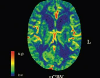
Although seizures are classically associated with abnormal electrical activity, recent work has suggested that there is also disrupted vascular activity following a seizure. Our lab has been involved in ground-breaking recent work that found significant and long-lasting blood flow reductions in the brain following a seizure. In humans, we have been able to detect blood flow reductions using arterial spin labelling MRI that last up to an hour following a seizure, and tend to co-localize with the region of the seizure onset. Due to the logistic constrains of obtaining an MRI within an hour window, we have also shown the same results using perfusion CT, which is faster, more readily available, and cheaper.

We have recently begun a small clinical trial that aims to determine whether commonly available medications (nifedipine and ibuprofen) can be used to prevent blood flow reductions following a seizure. We are currently working towards replicating rodent findings that demonstrate a relationship between blood flow reductions and cognitive and behavioural deficits following a seizure. Similarly, as part of the clinical trial, we aim to determine whether ibuprofen and nifedipine can prevent these deficits.

Click here to see our recent paper on posticital hypoperfusion and SUDEP (sudden unexpected death in epilepsy) or here to see our recent paper on localizing the seizure onset zone using postictal hypoperfusion.

Collaborators

We have ongoing collaborations with many outstanding researchers, including:

Dr. Bradley GoodyearDr. Cam TeskeyDr. Richard FrayneDr. Bruce PikeDr. Pierre LeVanDr. Julia JacobsDr. Colin JosephsonDr. Sam WiebeDr. Karl Martin KleinDr. Bijoy MenonDr. Ting Lee (Western University), Dr. Marc Lebel