Current Projects
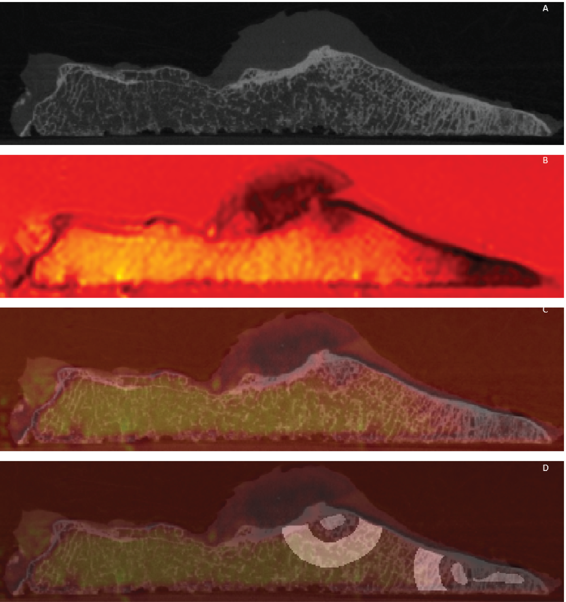
Determining the role of bone marrow lesions in knee osteoarthritis: Investigating high bone turnover in vivo
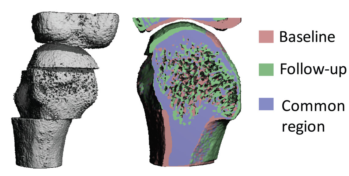
Knee osteoarthritis (OA) is a degenerative disease that causes significant morbidity and a loss of quality of life. Advances in treatment to slow or prevent joint degradation have been limited due to the lack of understanding of the timing of changes in joint tissues, and the poor association between pain and radiographical features of OA. Bone marrow lesions (BMLs) are one structural feature of OA that have been consistently linked to pain in knee OA. BMLs are known to be sites of high bone turnover and sclerosis, however, the cause and effect of this increased bone metabolism is not yet understood. In this study, we will use MRI to locate BMLs, and second-generation HR-pQCT to accurately visualize subchondral bone microarchitecture at 61 um resolution. Using image registration, we will quantify bone turnover rates directly in the regions containing BMLs.

Understanding Muscle Loss in Critical Care Patients: Opportunistic Evaluation of Muscle and Bone
Muscle loss can lead to weakness, fracture, and a reduced quality of life. In critical care patients, low muscle density is linked to higher mortality, and in survivors, symptoms of this muscle loss as well as bone loss can persist long-term. CT images taken of patients at admission and discharge can be analyzed retrospectively using a recently developed internal calibration method to evaluate muscle and bone health outcomes. Understanding the complexities of care and specific challenges that individuals experience could help indicate risk factors for bone and muscle loss, as well as better inform treatment options. This project aims to utilize these opportunistic clinical CT scans to validate the internal calibration method for muscle density analysis.

The relationship between sub-clinical inflammation and bony damage progression in rheumatoid arthritis
Rheumatoid arthritis (RA) is a chronic autoimmune condition targeting the synovium of the small joints of the hands, wrists, and feet, and in particular the 2nd and 3rd metacarpophalangeal (MCP) joints. Timely, target-driven therapeutic intervention with disease-modifying agents is currently the most effective mechanism to achieve clinical remission and prevent joint damage. Imaging plays an important role in evaluating disease onset, progression and treatment effectiveness. However, a disconnect between clinical remission and imaging remission has been demonstrated - some patients in sustained clinical remission still have evidence of active inflammation on ultrasound or MRI, and continue to have worsening radiographic damage scores. HR-pQCT is provides a more sensitive assessment of bone damage including bone microarchitecture, joint space width, as well as erosion number and size. We will determine what level of inflammation, assessed by MRI, is associated with bony damage progression, assessed by HR-pQCT.

Characterizing the role of trapeziometacarpal joint biomechanics on the development and progression of osteoarthritis in the thumb
Osteoarthritis (OA) in the hand affects millions of people worldwide, yet receives relatively less attention than other forms of OA. One of the commonly affected joints in the hand is the trapeziometacarpal joint of the thumb (TMC), resulting in increased pain and a decreased ability to perform activities of daily living. Abnormal biomechanics and loading patterns are a risk factor for the development and progression of OA in the hand. The ability to detect and quantify subtle changes during joint motion may aid in furthering our understanding of the relationship between joint structure and function in diseases such as OA. Dynamic CT is a relatively new technique in the musculoskeletal imaging field that utilizes three spatial dimensions as well as time to provide dynamic, three-dimensional (3D) representations of bone and joint biomechanics. This project aims to develop a fast, semi-automated processing pipeline for dynamic CT to quantify and visualize specific biomechanical outcome measures beyond what is available with static CT or motion capture technology.

