Small Vessel Disease Imaging

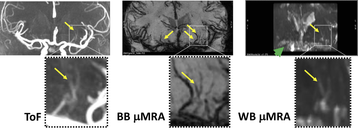
Comparison of experimental time-of-flight (ToF) black-blood (BB) and white-blood (WB) imaging techniques for examining smaller cerebral blood vessels.
We use MR imaging to study pathology in cerebral small vessel disease (cSVD).
Our methods include T1-weighted volumetric imaging (to look for brain atrophy), T2-weighted and FLAIR imaging (to look for white matter hyperintesities), arterial-spin labelling (ASL) perfusion imaging (to look for hypoperfusion), diffusion imaging (to examine key aspects of tissue integrity) quantitative susceptibility mapping (QSM, to look for iron deposition and microbleeds), and T1 and T2 relaxation time mapping (to examine subtle changes in tissue chemistry).
Over the past decade, we have played a significant role in the development of standards for imaging cSVD:
- Wardlaw JM, Smith EE, Biessels GJ, Cordonnier C, Fazekas F, Frayne R, et al. Neuroimaging standards for research into small vessel disease and its contribution to ageing and neurodegeneration. Lancet Neurol 2013; 12: 822-38. doi: 10.1016/S1474-4422(13)70124-8
and its 2023 update:
- Duering M, Biessels GJ, Brodtmann A, Chen C, Cordonnier C, de Leeuw FE, Debette S, Frayne R, et al. Neuroimaging standards for research into small vessel disease-advances since 2013. Lancet Neurol 2023; 22: 602-618. doi: 10.1016/S1474-4422(23)00131-X
In addition to exploring quantitative structural and functional measurements, we are also interested in developing clinically useful, high resolution MR angiography (µMRA) techniques to examine small arteries (<500 µm), such as the lenticulostriate and deep perforating arteries. Occlusion of these vessels have been implicated in cSVD. We are currently exploring the development of both white and black blood-based approaches (WB and BB, respectively).
Many of these activities occur in collaboration with Eric Smith of the Clinical Research Laboratory on Vascular Cognitive Impairment and Philip Barber of the Calgary Stroke Program.
Representative Publications
- MacDonald ME, Frayne R. Cerebrovascular MRI: a review of state-of-the-art approaches, methods and techniques. NMR Biomed. 2015; 28: 767-91.
- Munir M, Ursenbach J, Reid M, Gupta Sah R, Wang M, Sitaram A, Aftab A, Tariq S, Zamboni G, Griffanti L, Smith EE, Frayne R, Sajobi TT, Coutts SB, d'Esterre CD, Barber PA; Alzheimer's Disease Neuroimaging Initiative. Longitudinal brain atrophy rates in transient ischemic attack and minor ischemic stroke patients and cognitive profiles. Front Neurol. 2019; 10: 18. doi: 10.3389/fneur.2019.00018.
- McCreary CR, Beaudin AE, Subotic A, Zwiers AM, Alvarez A, Charlton A, Goodyear BG, Frayne R, Smith EE. Cross-sectional and longitudinal differences in peak skeletonized white matter mean diffusivity in cerebral amyloid angiopathy. Neuroimage Clin. 2020; 27:102280. doi: 10.1016/j.nicl.2020.102280.
- Zhu Z, Lebel RM, Bliesener Y, Acharya J, Frayne R, Nayak KS. Sparse precontrast T1 mapping for high-resolution whole-brain DCE-MRI. Magn Reson Med. 2021; 86: 2234-2249. doi: 10.1002/mrm.28849
- Reid M, Tadros GS, McDougall CC, Reaume N, McDougall B, Sah RG, Wang M, Smith EE, Frayne R, Coutts S, Sajobi T, Longman RS, d'Esterre CD, Barber P. Arterial spin labelling reveals multi-regional cerebral hypoperfusion in patients with transient ischemic attack that are unrelated to ischemia location: A proof-of-concept study. Cereb Circ Cogn Behav. 2023; 4: 100164. doi: 10.1016/j.cccb.2023.100164
- Sharma B, Beaudin AE, Cox E, Saad F, Nelles K, Gee M, Frayne R, Gobbi DG, Camicioli R, Smith EE, McCreary CR. Brain iron content in cerebral amyloid angiopathy using quantitative susceptibility mapping. Front Neurosci. 2023; 17: 1139988. doi: 10.3389/fnins.2023.1139988
Prospective Trainee Requirements
Interest in cerebrovascular disease with a degree (MSc preferred) in Biomedical, Computer or Electrical Engineering, or Physics. A good grasp of digital signal processing techniques and/or experience with numerical optimization/modelling methods beneficial. Must have good oral and written English communication skills.
