Neonatal Neuro-Critical Care Innovation in Education Lab
About the lab
The Neonatal Neuro-Critical Care Program under the umbrella of the ACH Neuro-Critical care Program has developed an innovation in Neonatal Neuro-Critical Care Education Lab. The lab has produced (all developed locally) cutting edge innovative simulators and models that have been used across the globe for clinical care application, quality improvement and research.
Vision:
A world where all trainees have access to innovative and affordable teaching modules to enhance knowledge and care for brain health.
Mission:
To create cutting edge, innovative teaching modules through collaboration with other specialties using the most recent available technology. Throughout our work in the lab we involved computer science students, bioengineering students, software engineers, 3D designers and mechanical engineering students in different projects. One of the computer science master's students successfully completed her master's degree on our head ultrasound simulator. Two students received a student summer grant and another has applied. If you are interested please contact us.
Cranial Ultrasound Simulation Model:
Computer-Based and Brain Phantoms:
This was developed initially in collaboration with the sonographic Clinical Assessment of the Newborn Program.
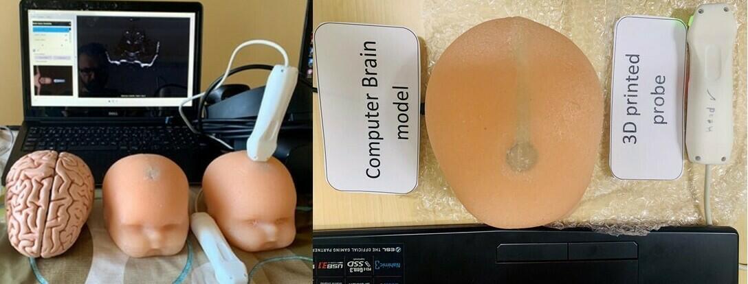
The cranial ultrasound simulators allow learners to learn and practice ultrasonography using a bank of images obtained from patients with normal cranial structures and multiple pathologies, and ultrasound-able 3D printed heads with landmark brain structures. The preloaded images can be selected using the computer program, while the mannequin with sensors embedded under the scalp, allows the probe to select corresponding image sequences to simulate real life scanning. The components include a laptop computer connected to a sensor within the probe. The purpose of these tools is to familiarize trainees with probe orientation, image optimization skills and ultrasound machine knobology.
The Cranial Ultrasound simulator was developed at the University of Calgary by the Neuro-Critical Care Program based at the Alberta Children’s Hospital in partnership with the SCAN group.
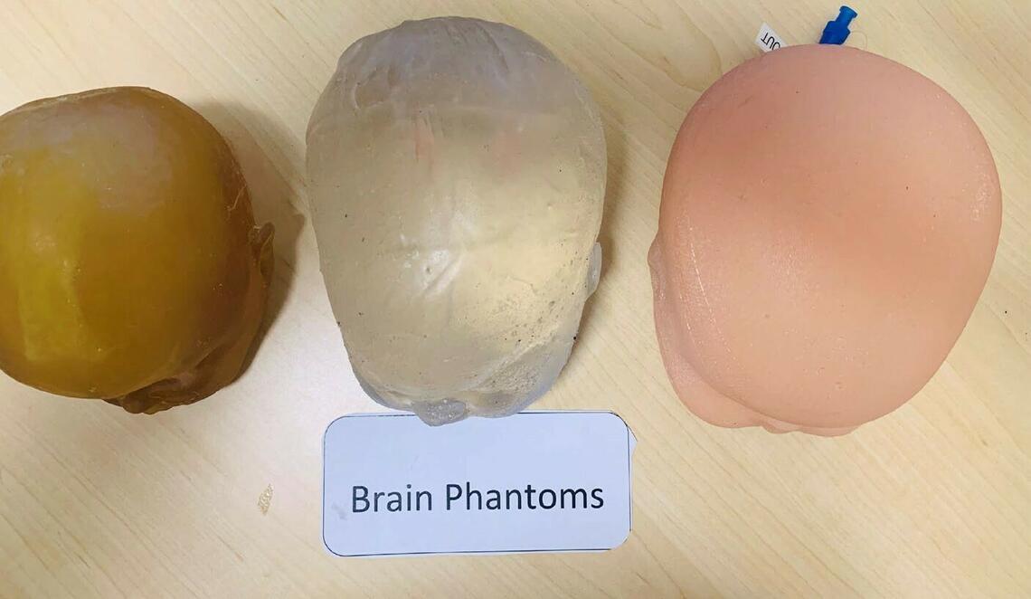
Brain Phantoms
Ultrasound-able 3D printed heads with simplified key internal brain structures (lateral ventricles, caudo-thalamic notch and choroid plexus).
Computer-Based Cranial Ultrasound Simulators
- 3D printed heads with pressure sensors at the locations of the anterior and mastoid fontanels.
- 3D printed probes with Arduino sensors inside them to interact with the sensors in the 3D printed heads.
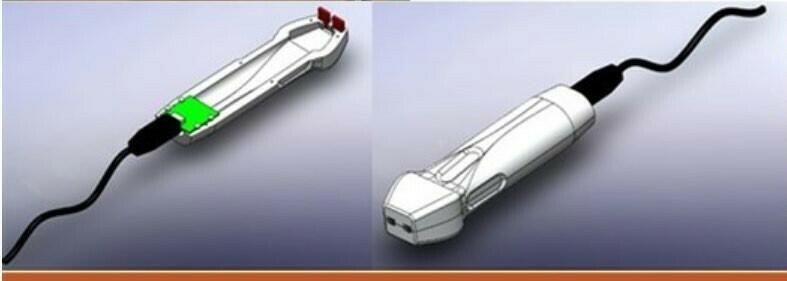
- Cranial ultrasonography software translates probe movements into images in a way that simulates a cranial ultrasonography study.
Implementation of The Models:
- The models have been used through conferences and hands-on workshops in several countries around the world (India, China, US, UK, Kuwait, Oman, Columbia and Quito) with more 1000 participants in total. We followed up with the centres after conferences and workshops organized by our group to help them build their programs and sustain the learned skills.
Clinical Skills and Education:
- The simulators have been integrated into our neonatology fellowship program. Currently all of our neonatology fellows get trained in neonatal cranial ultrasonography, compared to no training before we developed the models.
Quality improvement:
- The models have been used as a tool to standardize preterm brain injury definition by radiologists and neonatologists: two workshops were conducted in Calgary and Toronto with representatives from radiology and neonatology from each level-three centre in the country. The core group has become a taskforce which since created a consensus practice guideline to standardize the diagnosis and imaging in preterm infants across the country.
Research:
- We used the model in testing the efficacy of simulation-based learning in gaining and sustaining ultrasonography skills. We published the results in a peer reviewed journal.
- In the development, is a smart tracing of the lateral ventricle borders to measure surface area to compare it with traditional 2D measurements to better predict the ideal time for surgical intervention.
Future planning:
- Adding MRI cases side by side to the same US pathology to be sliced simultaneously while performing cranial ultrasound
- Create interactive interface (quizzes and labeled anatomy and pathology) to help trainees self-learn using the simulator
- Allow physicians to add their own cases and create center specific libraries
- Create brain phantoms with corpus callosum , cerebellum, and third ventricle
- Adding cerebral arteries doppler functionality
Cerebral Arteries Doppler Model:
This model simulates the anterior cerebral artery embedded in an ultrasound-able brain phantom. The artery is connected to a peristaltic pump to generate the flow. A 20 cc syringe is used to manually generate the pulsation. Participants can generate 2D images of the anterior cerebral artery, learn doppler acquisition skills (color doppler, pulse wave doppler, and measurement of resistive and pulsatility indices).
Implementation of the Models:
The model was used in 2 workshops in Kuwait and Banff with excellent feedback.
Future Planning:
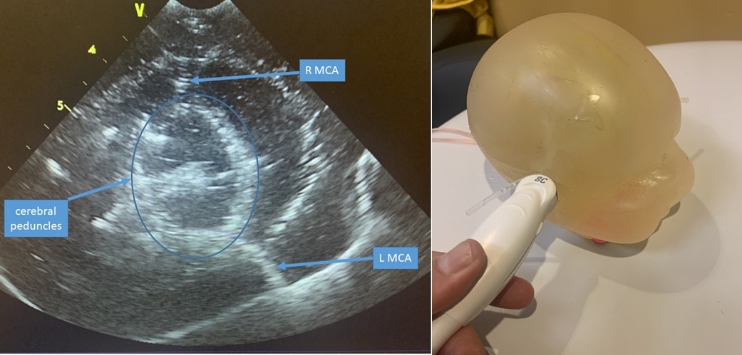
- Development of middle cerebral arteries and cerebral peduncle.
- Create smaller and more flexible vessel to generate more realistic pulsatility.
- Purchase a pulsatile pump to mimic cardiac stroke.
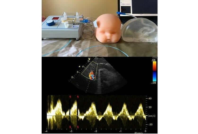
MCA model:
PHVD measurement and Reservoir Tapping Model:
As the surgical intervention for post hemorrhagic ventricular dilatation ( PHVD) is becoming rare, maintaining competency in taping the ventricular reservoir is a challenge. We developed a “just-in-time” teaching model which includes:
Implementation of the Model:
- The model is used as a “just in time” teaching tool each time we face a case of reservoir insertion.
- The model was used in 2 workshops: at the Pediatric Academic Society and the American Academy of Pediatrics District VIII section of Neonatal- Perinatal Medicine.
Future Planning:
To develop hydrocephalus brain phantoms with different ventricular sizes
Smart Phone Application for Preterm Brain Injury Diagnosis:
We converted the consensus view point that we developed between neonatology and Radiology into a smartphones application and made it free on google play and iPhone to improve compliance and decrease variability.
Neonatal Neurological Examination Teaching Simulators and Modules:
The purpose of these set of simulation models and teaching module was to improve identification of infants suffered from perinatal asphyxia and eligible for therapeutic hypothermia due to short window of opportunity to start the treatment.

Targeted Neurological Examination Teaching Videos:
A formal consent was taking from families and full neurological examination videos were recorded for normal and abnormal newborns. The videos were then edited and integrated into one targeted neurological examination to improve HIE identification.
Cooling calculator for Android devices
Cooling calculator for Apple devices
Coming soon...
Cooling calculator for PCs
Targeted Neurological Examination Virtual Reality Module:
Recorded videos were converted in animation scenarios then used to create a virtual reality module in which trainees can observe and perform targeted neurological examination.
Neurological Examination Mannequins:
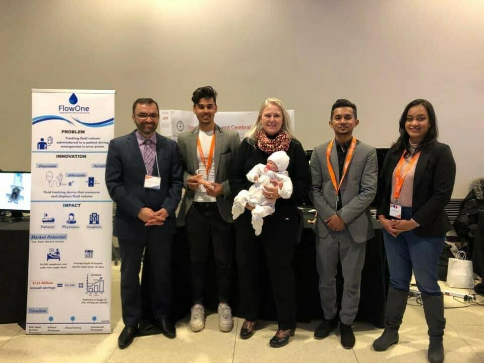
In collaboration with a group with computer science, mechanical engineers, and bioengineers, a neurological examination model was developed to simulate the tone assessment and posture.
The project won the best in clinical category at the 2019 health hack competition.
Clinical Skills and Education:
The combined package of teaching modules and simulators were used in the Southern Alberta Neonatal Outreach program where we vested all level I and II referring centres and conducted sessions on improving HIE identification.
The models were used at 2 workshops in Kuwait and Nursing neonatal Neuro-Critical care workshop in Calgary.
Impact:
After the implementation of theses teaching simulators and modules the rate of missed cases decrease significantly from 28% to 7% without increasing our total annual number.
Research:
A research study was designed and approved by the ethics committee to compare traditional methods in teaching neurological examination to the simulation-based method.
Neonatal Electro-Encephalogram (EEG) Teaching Modules and Simulator:
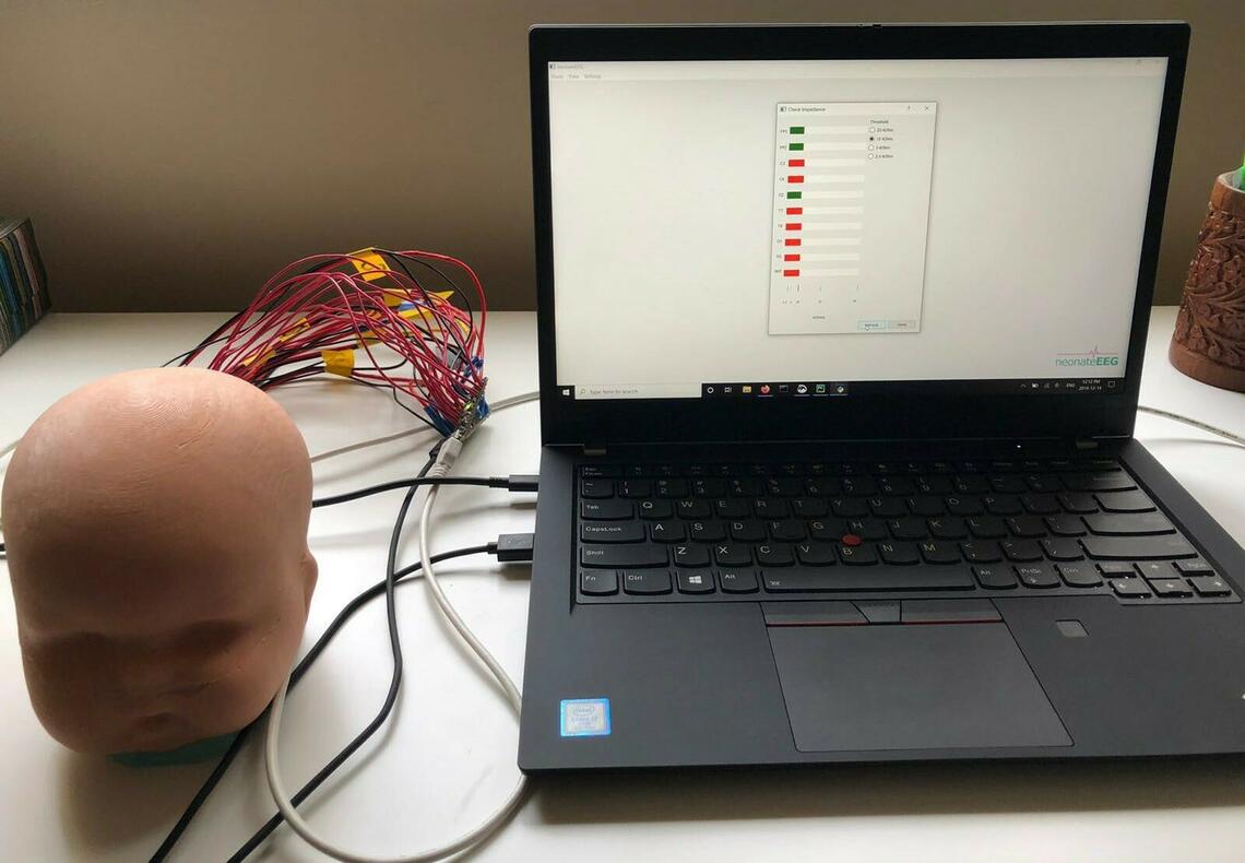
Teaching videos were created to demonstrate how to place EEG electrodes and generate a good quality signal with "step by step - how-to" tutorials on starting an EEG study and troubleshooting.
In collaboration with computer science, mechanical engineers, software engineers, and mechanical engineers an EEG simulator was developed to simulate EEG set up.
Clinical impact:
Using all teaching modules (teaching videos, documents, small group training, workshops, and recently the simulator) we trained NICU nurses to set up EEG studies which helped in the establishment of our neuro-monitoring in the NICU. Having NICU nurses initiate EEG studies significantly decreased time to initiate EEG studies. Establishing neuro-monitoring in the NICU resulted in significant reduction in anti-seizure medication use and improved seizure diagnosis.
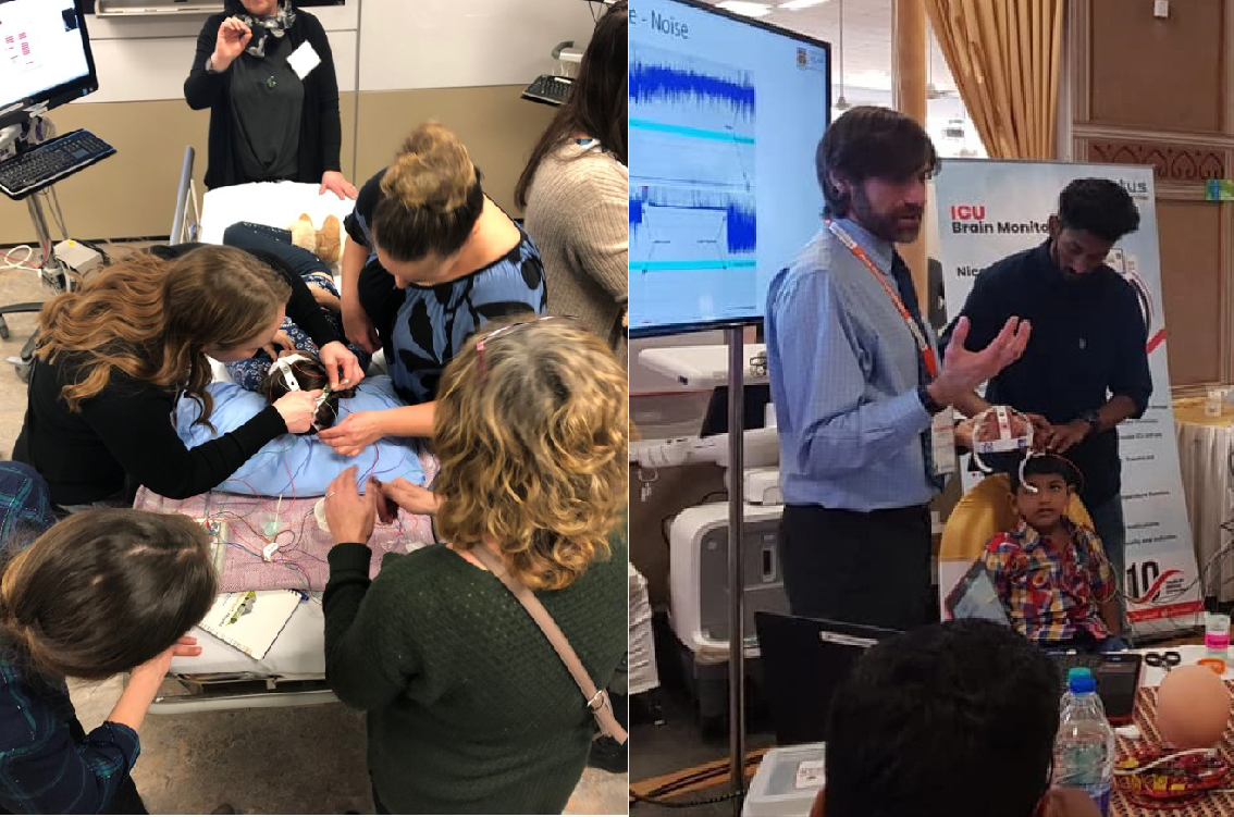
Teaching EEG Set up Using Simulation and Live Children:
The EEG and neuro exam simulators were used at our local, national and international workshops with our own children volunteering as simulation models.
Research:
A research study was designed and approved by the ethics committee to compare traditional methods in teaching EEG set up to the simulation-based method.
The goal of educational modules and applications is to serve as a resource for trainees , nurses and physicians interested in Neonatal Neuro-Critical Care, However, it must be understood that it is in no way intended to provide recommendations for clinical decision making and family counselling. The decision making tools and educational materials included do not, in any way, constitute a diagnostic interpretation or clinical recommendations for patient care on their own . The authors and their institutions including the Calgary Neonatal Neuro-Critical care program are not responsible for any decision making based on findings from this application. All rights reserved. No part of these teaching modules may be reproduced or modified in any way without the prior written permission of the Calgary Neonatal Neuro-Critical Care program. Our team is committed to providing safe courses for participants. We comply with Canadian Federal and Provincial privacy laws and regulations including the Personal Information and Electronic Documents Act. Images and videos should be de-identified and formal consent should be taken from families. Images, details, and investigations shared and legal implications and responsibility lie with the person who is sharing. The purpose of these courses is educational. They don't give physicians, trainees, nurses or participants any privileges or generate any kind of licensing. Team members, their institutions or programs are not responsible in any way for clinical decisions or diagnoses been made based on skills gained using these courses.
These modules were developed in collaboration with and support from Pediatric Neuro-Critical Care and Sonographic Clinical Assessment of the Newborn (SCAN) programs in Calgary. The HIE Surveillance and outreach programs, and innovation lab are supported by the Calgary Health foundation. Education modules and innovation lab have received an educational grant and in-king support from Alberta Children Hospital Research institute (ACHRI), Alberta Children Hospital foundation and Pediatric Neuro-Critical Care program. None of Scientific committee members or speakers have conflict of interest to disclose.
Learn about Calgary Health Foundation
Scientific faculty and speakers (in alphabetical order)
None of Scientific committee members or speakers have conflict of interest to disclose
- Dr. Ayman Abou Mehrem, MD, University of Calgary
- Dr. Juan Pablo Appendino, MD, University of Calgary
- Dr. Zarina Assis, MD, University of Calgary
- Dr. Luis Bello-Espinoza, MD, University of Calgary
- Dr. Amina Benlamri, MD, University of Calgary
- Dr. Susanne Benseler, MD, University of Calgary
- Naureen Bhamani, Alberta Children Hospital , Calgary
- Dr. Marie-Anne Brundler, MD, University of Calgary
- Dr. Stefani Doucette, MD, University of Calgary
- Dr. Michael Esser, MD, University of Calgary
- Leah Foster, NP, ALbertal Children Hospital , Calgary
- Dr. Elsa Fiedrich, MD, University of Calgary
- Dr. Leonora Hendson, MD, University of Calgary
- Dr. Alice Ho , MD, University of Calgary
- Dr. Adam Kirton , MD, University of Calgary
- Silvia Kozlik, Alberta Children Hospital , Calgary
- Dr. Lara Leijser, MD, University of Calgary
- Dr. Julia Jacobs-LeVan, MD, University of Calgary
- Jan Lind , RN , Alberta Children Hospital
- Dr. Aleksandra Mineyko, MD, University of Calgary
- Dr. Khorshid Mohammad, MD, University of Calgary
- Dr. Prashanth Murthy, MD, University of Calgary
- Norma Oliver, RN , Alberta Children Hospital
- Dr. Eric Payne, MD, University of Calgary
- Dr. Morris Scantlebury, MD, University of Calgary
- Dr. James Scott , MD, University of Calgary
- Dr. Amelie Stritzke, MD, University of Calgary
- Dr. Sumesh Thomas, MD, University of Calgary
- Dr. Xing-Chang Wei, MD, University of Calgary
- Dr. Hussein Zein, MD, University of Calgary
Student Volunteers
- CME application
- Yasmina Menhem
- Animation and interactive quizes
- Kiaksar Mohammad

