Kindly sponsored by:



Hosted and organised by the Advanced Imaging and Microscopy Network (AIM), the competition celebrates microscopy excellence and promotes colour blind friendly images. Showcase your research by submitting biology-related images captured on any optical microscope across the University of Calgary!
Click below for more information about the competition and submission process.

2022 Winners
Grand Prize Winner
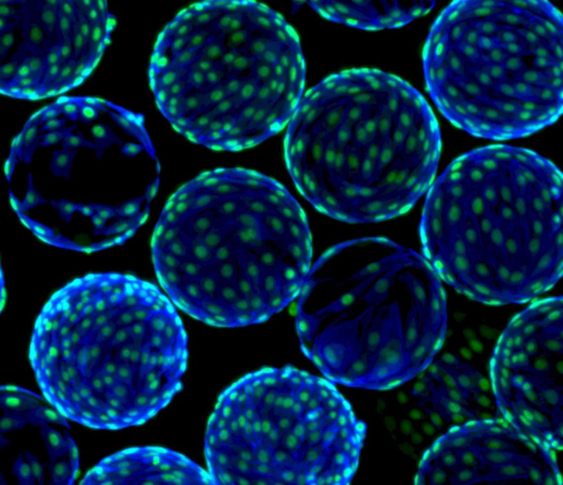
This image shows cord blood mesenchymal stem cells grown on microcarriers (small plastic beads ~200um in diameter) in a bioreactor. The green is a nuclei stain and the blue is an actin stain.
Erin Roberts
ACHRI First Prize
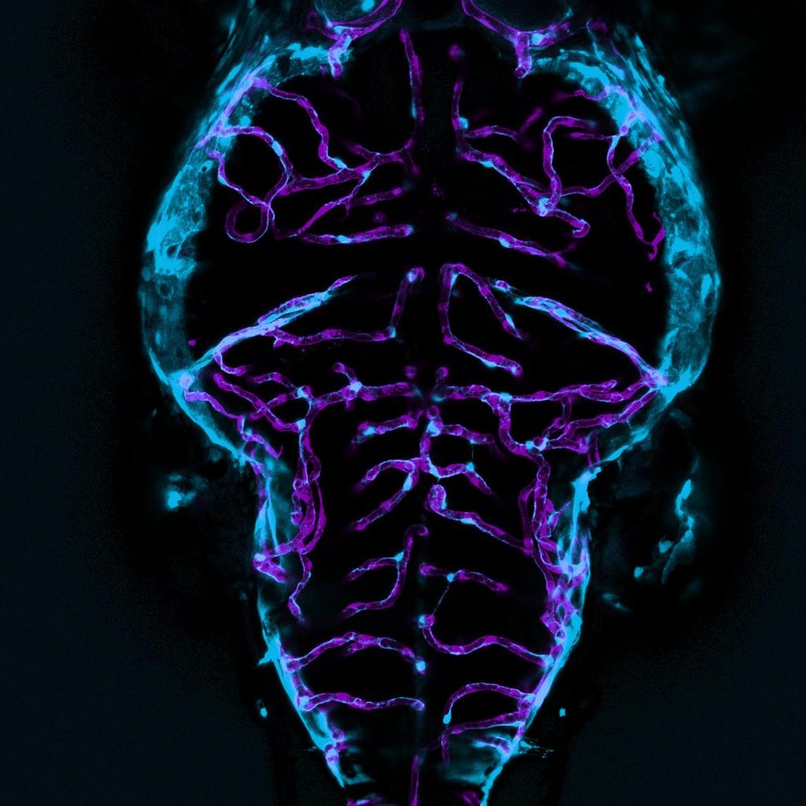
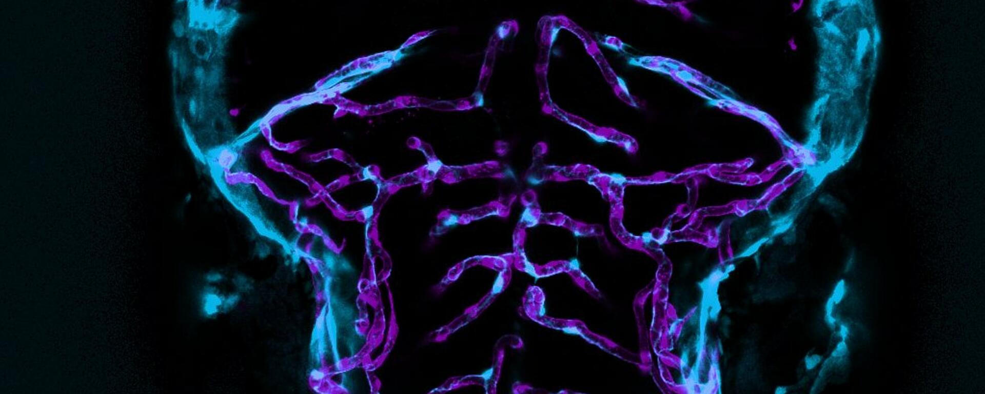
Zebrafish brain vasculature depicting endothelial cells (purple) and pericytes (cyan). Transgene labeling - Pericytes with pdgfrB: GFP Endothelial cells with kdrl: mCherry
Cynthia Ufuoma Adjekukor
CMF Category First Prize
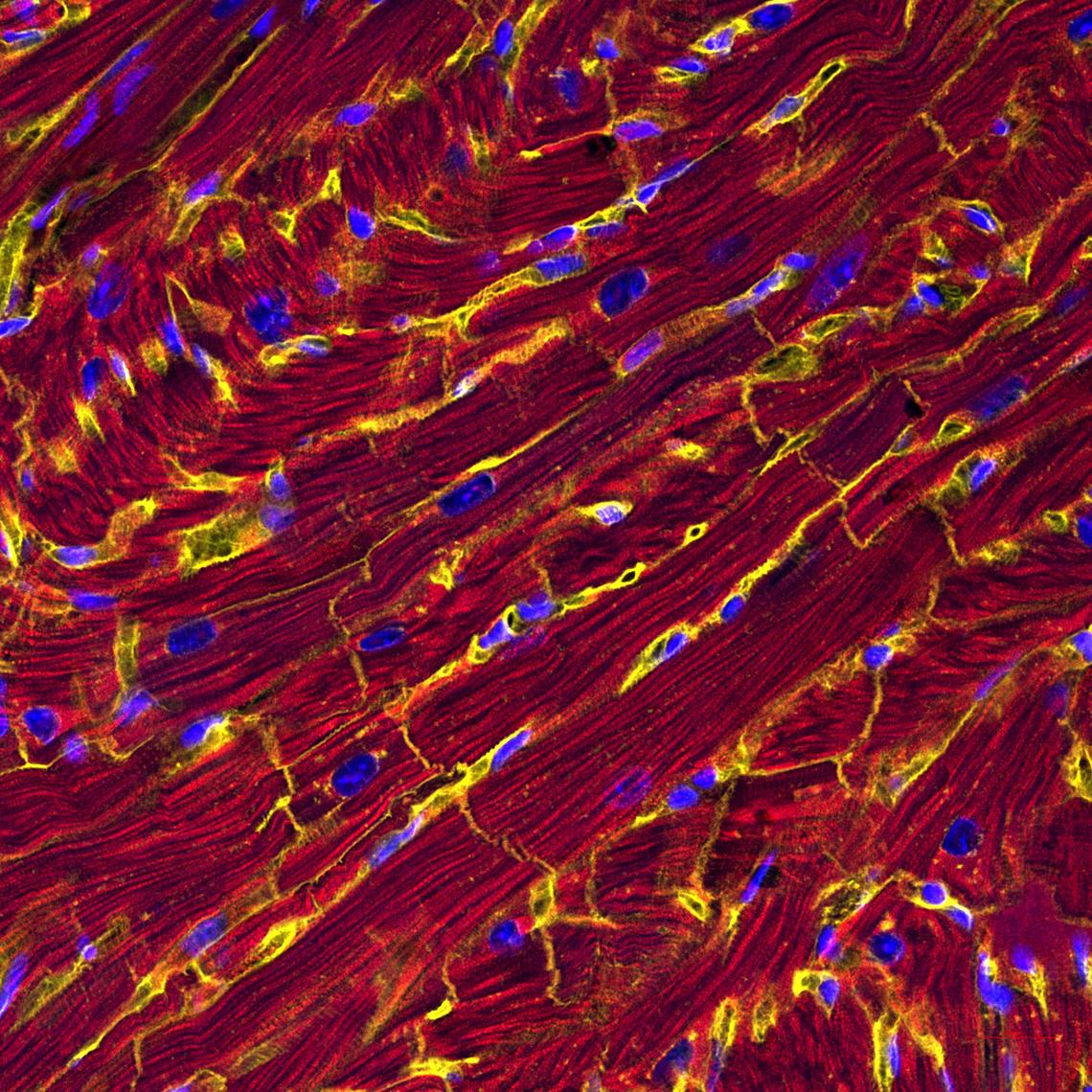
Mouse cardiomyocytes: Mouse heart cells probed for cell membrane (yellow), contractile structure and volume (red), and nuclei (blue) to study the effect of ING5 on cardiac function and recovery.
Hamed Hojjat
LCI Category First Prize
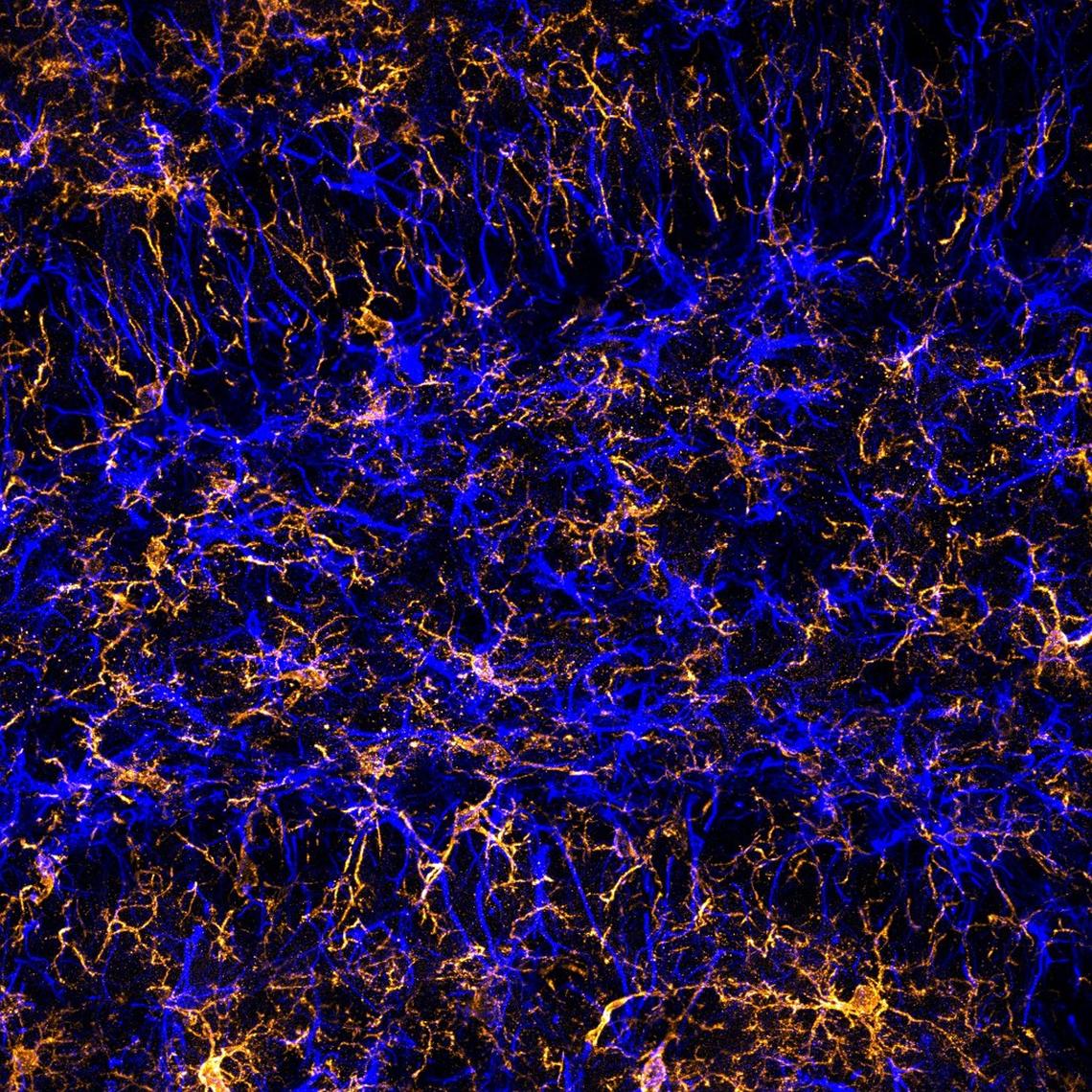
Microglia labeled with IBA1 (orange) and glia labeled with GFAP (blue) in the dentate gyrus region of the mouse brain.
Laurie Wallace
Artistic Catergory First Prize
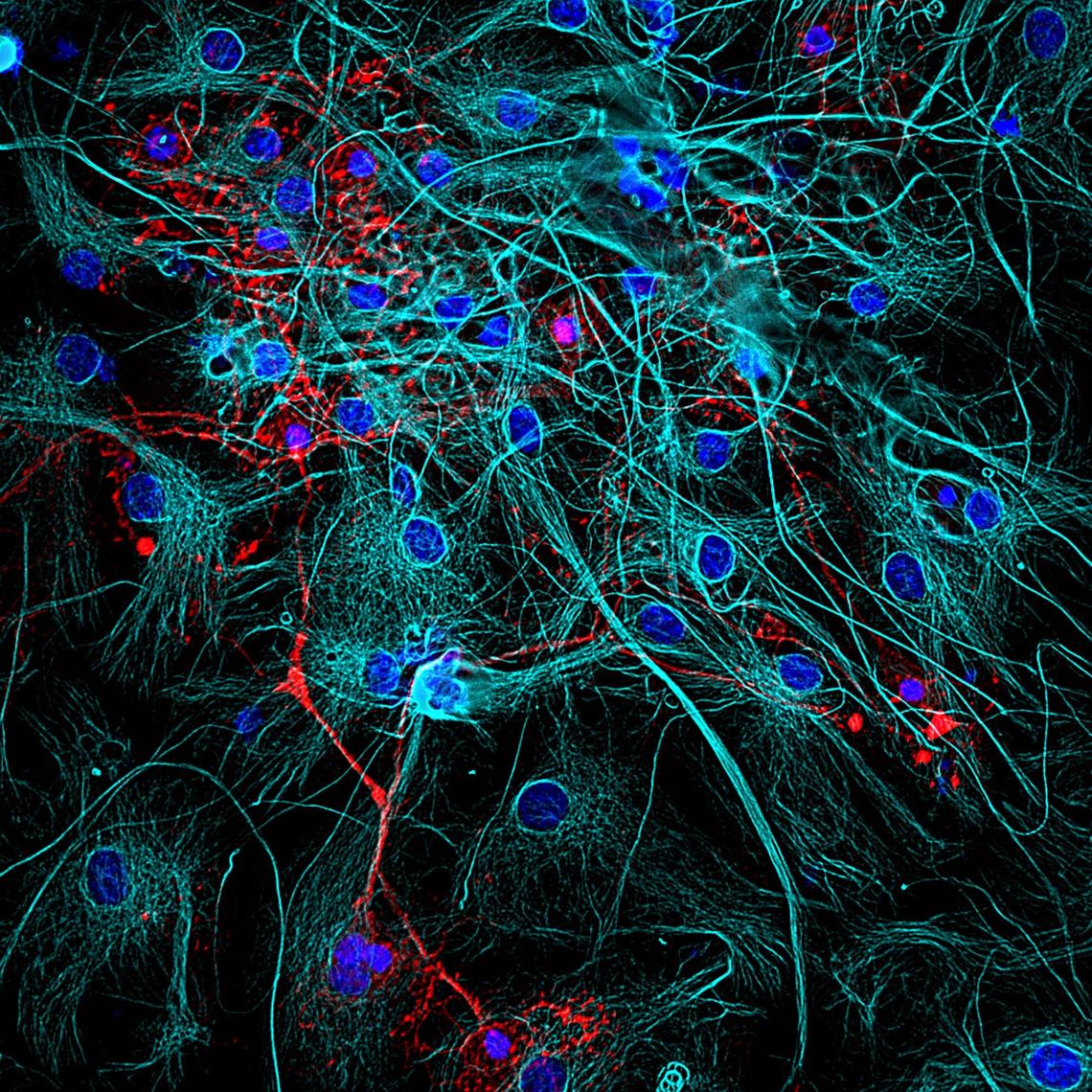
Astrocytes & Oligodendrocytes. The image shows a group of oligodendrocytes (Red) and Astrocytes (Cyan) in an extensive web of countless connections, highlighting a miniature of the delicate wiring of mammalian brain
Hiba Omairi
HBIAMP Category First Prize
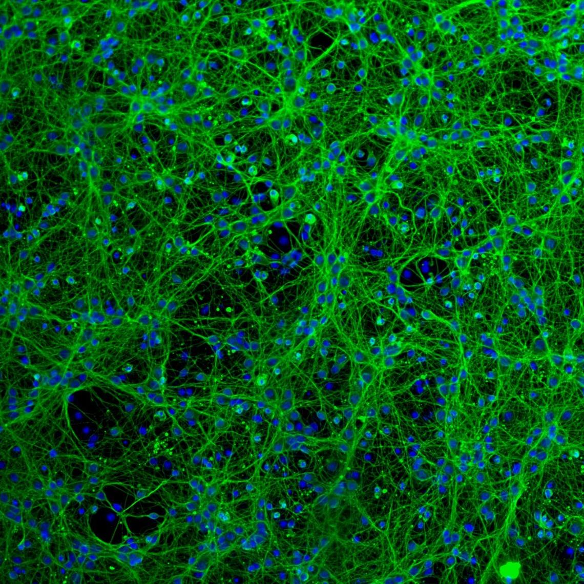
Embryonic mouse neurons were allowed to grow and form processes in a media culture for 72 hours without any stimulations. The cells were then immunofluorescently stained with antibodies to visualize their structure.
Dorsa Moezzi
Open Category Winner
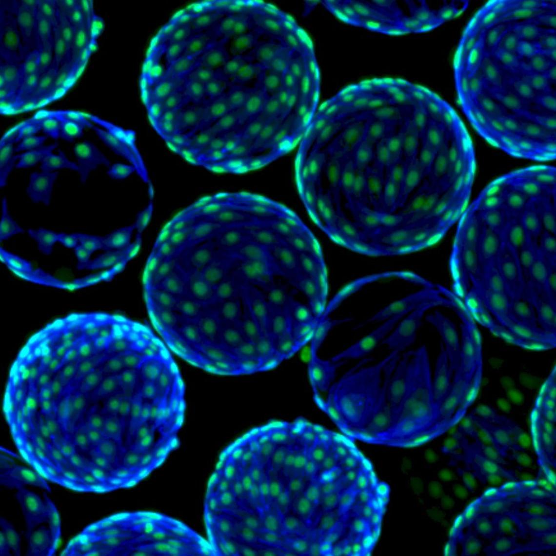
This image shows cord blood mesenchymal stem cells grown on microcarriers (small plastic beads ~200um in diameter) in a bioreactor. The green is a nuclei stain and the blue is an actin stain.
Erin Roberts

