Kindly Sponsored By:
Coming soon.
Hosted and organized by the Advanced Imaging and Microscopy (AIM) Network, the competition celebrates microscopy excellence and promotes colour blind friendly images. Showcase your research by submitting biology-related images captured on any optical microscope across the University of Calgary!
Striking Image Competition - Information and Submission
Rules, Criteria, Prizes, and Resources
Competition Information
Please read the following information before making your submission.
Submission Deadline: November 7, 2025
Maximum one entry per category per person.
All entries must:
- Be from biology-related research
- Have been acquired on an optical microscope at the University of Calgary
- Be colour blind friendly (see Resources section)
- Be made via our submission form
- Be accompanied by a 300-character lay summary (see Resources section
- Not contain embedded text (i.e. arrows, scale bars, annotations)
- Not contain multiple panels
- Not have been post-processed unless limited and adequately justified and described
File requirements:
- Submitted images must be in .TIFF format and limited to 100MB in size.
- Submitted videos are limited to 30 seconds.
Submitting an image or video implies that you (the submitter):
- Hold the rights to this image in terms of intellectual property and/or have obtained all necessary permissions to do so.
- Give permission to the Striking Image Competition organizers to use your name and image in connection with the contest. (Note: You are not giving up IP of this image to us or any other party. Files will be retained by the Striking Image Competition organizers.
Failure to comply with these rules will result in disqualification.
- Single Cell/Subcellular Images
- Tissue/Organ/Whole Body Images
- Lightsheet Images
- Videos
- Technical excellence (25%): focus, resolution, contrast/SNR, dynamic range, saturation (unless required to highlight dark features), depth-of-field, use of light; how demanding is it to acquire this image?
- Artistic merit (25%): design (balance, proportion, framing), use of colour/shape/space, uniqueness, impact on viewer, composition (rhythm, patterns)
- Scientific merit & communication (25%): lay summary (see Resources section) explaining the nature and/or the significance of the image/research within a broad context
- Colour blind friendliness (25%): how colour blind friendly (see Resources section) is the image?
The top THREE submissions in each category based on the judging criteria will be awarded prizes.
NB: Organizers reserve the right to modify the number of winners based on number of submissions and other factors.
About Colour Blindness & Colour Blind Friendly Images
- Colour Blind Awareness: Stats, Causes, Diagnosis, Types
- Considering colour blindness in manuscripts
- ImageJ tool to simulate colour blindness
About Lay Summaries
- Lay summary guidelines and examples for simplifying some scientific terms
- Example of 300-character lay summary (modified from Nikon Small World)
Trichome (fine outgrowths), stomata (small pores), and vessels of a southern live oak leaf. All three are essential to plant life: trichomes (white) protect against extreme weather and insects; stomata (purple) regulate the flow of gases in a plant; vessels (cyan) transport water throughout the leaf.
Submission Form
To submit an entry your must be a member of the University of Calgary with a valid @ucalgary.ca email address.
Submission form is open from September 15th to November 7th.
2024 Winners
First Prize
Second Prize
Shared Third Prize
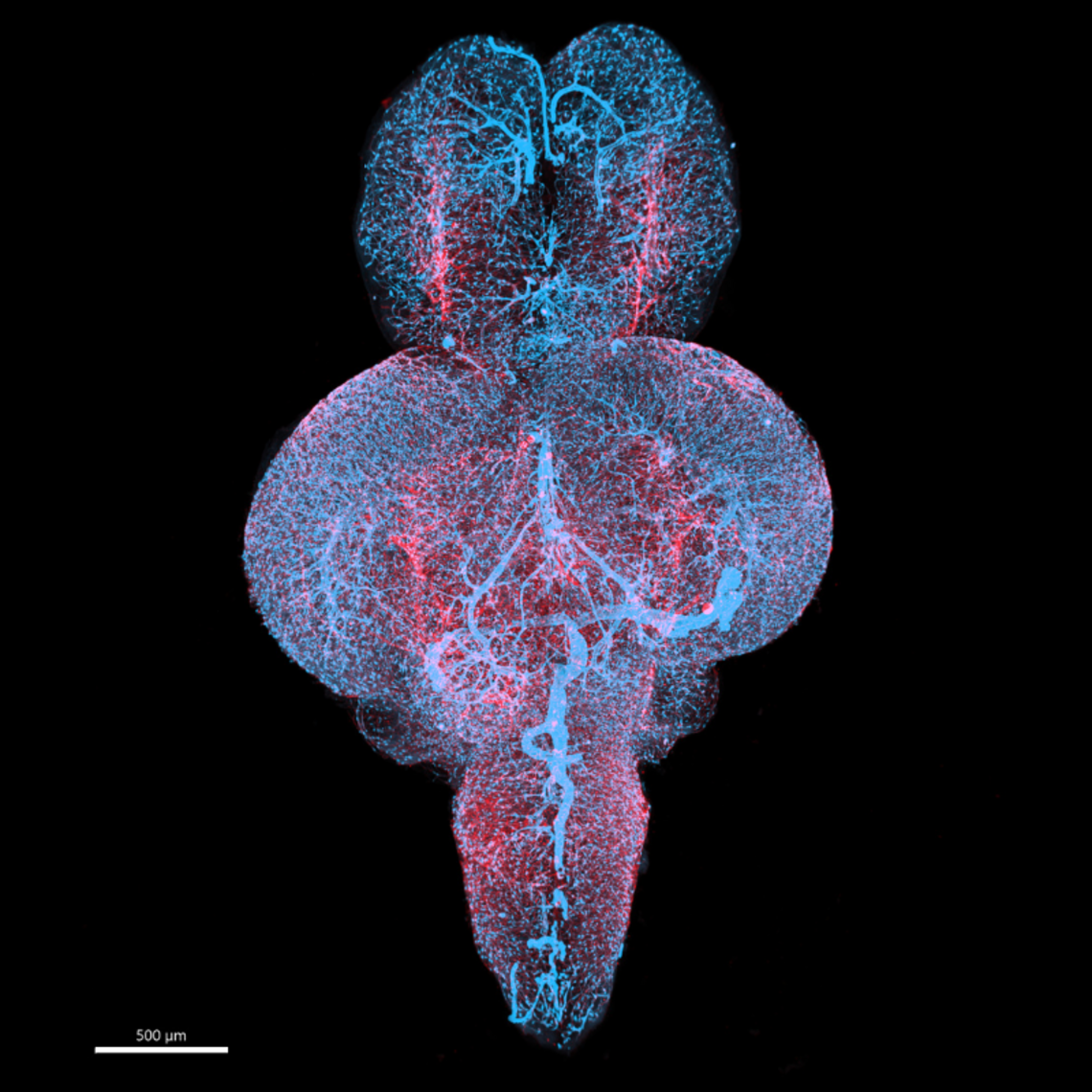
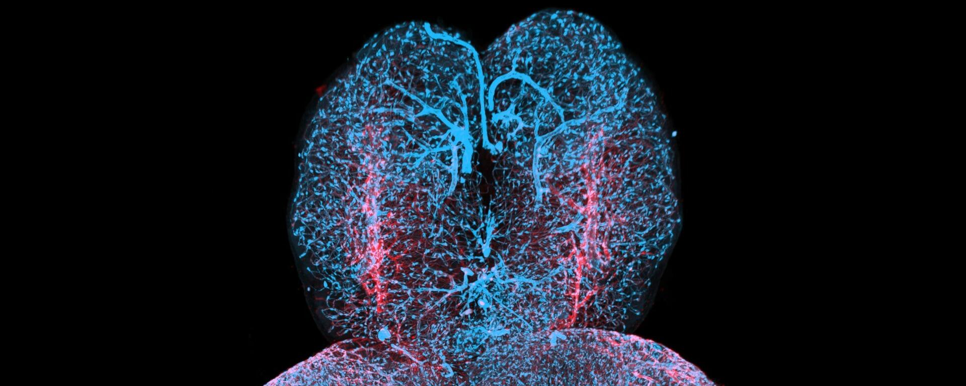
Pericytes (blue: "vascular support cells") and blood vessels (red) in a whole adult zebrafish brain. Pericytes are essential for vascular stability in the brain and defects in these cells and their coverage can underly vascular disease.
Merry Faye Graff
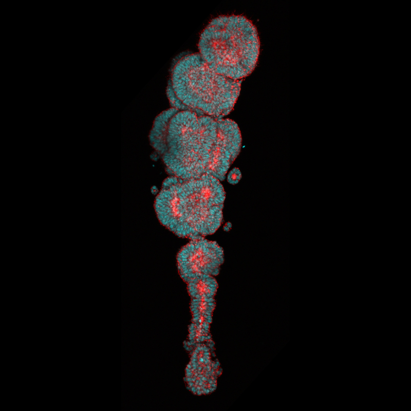
The image shows an organoid, which is simplified version of an organ grown from stem cells in a lab that can be used to understand its development and disease. This particular organoid represents a human "somite-forming" organoid.
Pranav Saligrama Ramesh
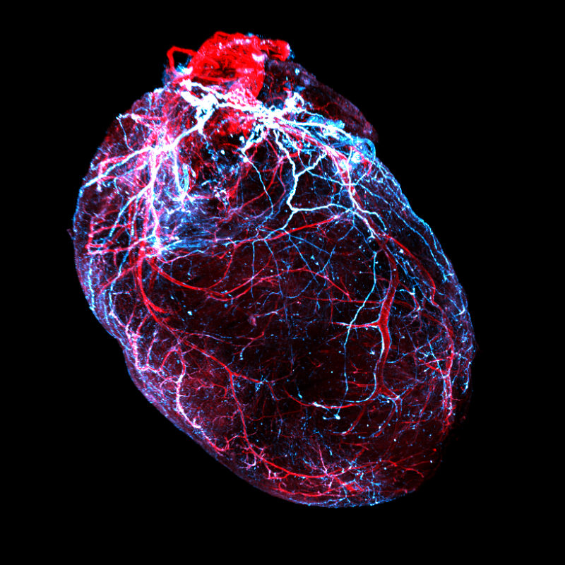
This image shows a transparent mouse heart, with the nerves in blue and blood vessels in red. We found that after spinal cord injury, nerve fibers in the heart are lost, leading to heart disease.
Jordan Lee
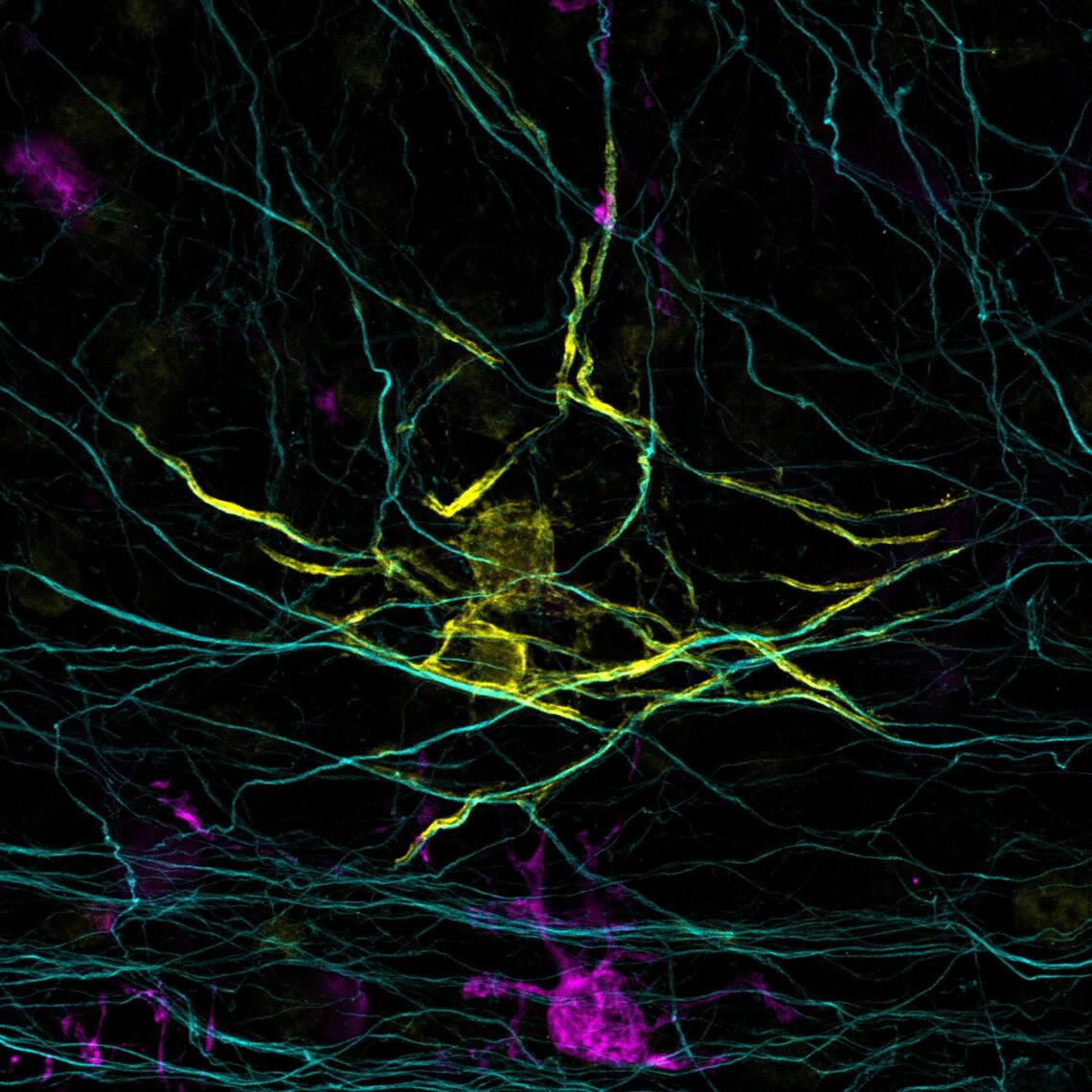
Your brain contains a large network of cables, over which electrical impulses are constantly exchanged. Oligodendrocytes wrap these cables in an insulating layer of myelin, speeding up brain signals by up to 100-fold. Here, you see an oligodendrocyte at work. Notice the pink cell below? That’s a microglia, one of the brain’s inspectors, stopping by.
Rianne Gorter
Honourable Mentions
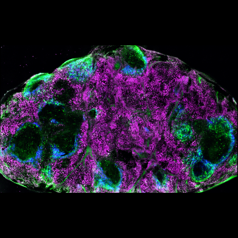
Oncolytic virus infection of the spleen drives anticancer immunity. F4/80+ macrophages are shown in magenta, CD169+ macrophages are shown in blue, infected cells are shown in green.
Victor Naumenko
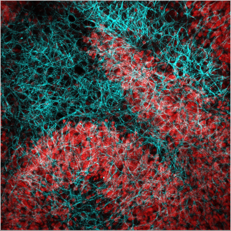
A sample of tissue from a mouse's cerebellum. I'm using this brain slice to study a new protein to see if it harms cells. By testing on this tissue, I can observe directly whether the protein is toxic and damages cells in a living-like environment.
Atefeh Rayatpour

